how do they x ray babies hips
You will go in the room with him he will need to be stripped from the waist down they will take x-rays of him flat on his back legs dead straight and together you wil be able to hold him in this position then an x-ray of his still on his back with his knees bent facing outwards and the soles of his feet put together he will be fine its not traumatic at all you will. Other bone tumors and tumor-like lesions such as eosinophilic granuloma may also be the underlying cause of a painful hip.

Pin On X Rays
The scan usually takes about 20 minutes.

. If a physical exam an ultrasound or an X-ray confirm a diagnosis your pediatrician will likely refer you to a pediatric orthopedic specialist for continued care and treatment. X rays CT scans and magnetic resonance imaging MRI scans may also be used. Most children do not need surgery but for those who do an arthrogram x-ray dye injected into the hip joint at the beginning of the surgery can help the surgeon decide exactly what needs to be corrected.
Youll be asked to partly undress your baby and take off their diaper for the test. Subsequent x-rays will track the hip joints progress. Up to 10 cash back Dog X-rays usually start around 200 and increase from there depending on how many images are needed.
For imaging assessment of developmental dysplasia of the hip ultrasound is the modality of choice prior to the ossification of the proximal femoral epiphysis. Two tests are performed called the Barlow and Ortolani tests to examine the function of the hip joints. Treatment for newborns A baby born with a dislocated hip can be successfully treated with a Pavlik harness.
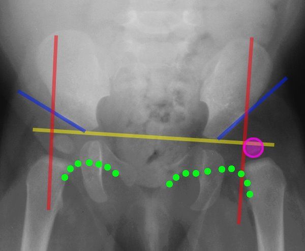
In Alaska I had like 5-10 infant chest X-rays a day and that thing made my job 100 percent easier. The most useful lines and angles that can be drawn in the pediatric pelvis assessing DDH are as follows. For some reason the left hip is said to be more frequently affected 4.
This image shows the soft tissues and the bones of the pelvis and hip joints. Usually both hips are scanned. The babys legs have differences in their lengths or appearances.
How do they X-ray babies hips. Around 6 months of age enough bone is present in an infant hip to make an X-ray more accurate than ultrasound. Ultrasounds are the diagnostic method of choice for infants under 6 months of age.
Because they spin around the body taking multiple images CT scans can deliver radiation doses that are up to 200 times higher than an average chest X. Each hip should be evaluated from the side and front view and many times images of the hip in a flexed or extended view are needed to assess if the hip joint is. Your baby was born in the breech position after 28 weeks of pregnancy.
However many more go undiagnosed as it may be too mild to even detect. The hip ultrasound will show the healthcare provider the position and shape of the hip joint. Radiographers appear to like it.
During the examination an X-ray machine sends a beam of radiation through the pelvic bones and hip joints and an image is recorded on a computer or special film. After around 4 to 6 months of age X-rays are the preferred method for evaluating and monitoring hip dysplasia. The doctor hears or feels a hip click when moving the infants thigh outward during a routine checkup.
This device holds the joint in place while the babys skeleton grows and matures. A Hilgenreiner line connects the inferior tips of the iliac bones at the triradiate cartilage. How do they xray babies hips.
Those things work great one person wrote on Reddit. An X-ray does not show the bones in a young baby until at least 6 months of age and therefore a hip ultrasound is preferred. X-ray imaging shows a small oval lytic lesion which may be obscured by the dense bony reaction surrounding it.
During treatment x-rays can reveal the progress of the hip as it improves. The purpose of this study is to report US results and follow-up of. The X-ray image is black and white.
Once there is a significant ossification then an x-ray examination is required. It is often the first type of imaging used to identify sources of pain evaluate traumatic injuries and locate a foreign body. When should I order an X-ray rather than an ultrasound to diagnose a musculoskeletal problem in an infant.
An X-ray of the pelvis focuses specifically on the area between your hips that holds many of your reproductive and digestive organs. Chest x-ray is the most commonly used imaging exam for evaluating the chest. X-rays are used throughout the body.
This image shows the soft tissues and. For example chest X-rays and hip X-rays require multiple images. A hip click can be felt by the examiner when the hip joints may not have formed normally.
During the examination an X-ray machine sends a beam of radiation through the pelvic bones and hip joints and an image is recorded on a computer or special film. It is estimated by the Center for Disease Control CDC that 1-2 of every 1000 babies have hip dysplasia. Your baby will be placed on a table on their back or side.
In babies with hip dysplasia the joint has not formed normally and the hips are prone to moving in and out of joint. Pediatricians do often check for hip problems in babies and hip dysplasia is the most common hip developmental deformity in children. MRI shows the nidus.
This line is used to measure the acetabular angle and as a reference for Perkin line. X-ray exams are used to help diagnose a wide variety of injuries and illnesses in children. How is hip dysplasia treated in babies.
If a hip click is felt your healthcare provider will usually obtain a hip ultrasound to assess the hip joint. If you have had twins or multiples and 1 of the babies has any of these risk factors each baby should have an ultrasound scan of their hips by the time theyre 4 to 6 weeks old. Sometimes a babys hip stabilises on its own before the scan is due but they should still be.
Because of the risk of developmental dysplasia of the hip in infants born breech-despite a normal physical exam-the American Academy of Pediatrics AAP guidelines recommend ultrasound US hip imaging at 6 weeks of age for breech females and optional imaging for breech males. This will require clear instructions for the parents to follow so that they do not allow rotation of the childs pelvis or motion artifact from kicking if the parent is accompanying the child by holding them in position whilst the parent puts on a lead gown it is the radiographers responsibility to ensure the baby does not roll off the x-ray.

Leerburg The Importance Of Good Positioning On Canine Hip X Rays Canine Hips Mastiffs

Basic Information About Dog Hip Dysplasia Paperblog Dog Hip Dysplasia Hip Dysplasia Canine Hip Dysplasia

Radiology Radiologic Imaging Signs List Collection Illustrated Cases Xray X Ray Gi Gu Chest Th Avascular Necrosis Radiology Avascular Necrosis Hip

Severe Hip Dysplasia In A Boxer The Red Arrows Are Pointing To The Over Growth Of Bone At The Femoral Neck Head T Shades Of Grey Hip Dysplasia Animal Heads
Lines Of The Hip Pediatrics Pediatrics Pediatric Nurse Practitioner Pediatric Radiology

Pin On Ortho

Uk Professor Says Swaddling Epidemic Gives Babies Clicky Hips Daily Mail Online Hips Professor Baby Swaddle

X Ray Image Of Child Swallowed The Coins For A Medical Diagnosis Medicine Pictures Children Images X Ray Images

Causes Of Ddh Hip Dysplasia Baby Developmental Dysplasia Of The Hip Baby Wearing

Pin On Spectacular Photography

23 X Rays Of Pregnant Animal Bellies That We Can T Decide Are Cute Or Creepy Pregnant Cat Pregnant X Ray

Hip Elbow Dysplasia Genetic Or Environmental Things To Ponder Hip Dysplasia Hips Canine Hip Dysplasia

Lower Limb Radiographs Anatomy And Physiology Anatomy Sacroiliac Joint

Hip Dysplasia In Adolescents And Young Adults Hss Hipproblems Hip Dysplasia Hip Problems Hips

Diagnosis Prevention And Management Of Canine Hip Dysplasia A Revie Vmrr Canine Hip Dysplasia Diagnostic Imaging Total Hip Replacement

Pin On Teach

Legg Calve Perthes Disease Radiology Nursing Radiology Calves
Avascular Necrosis Avascular Necrosis Juvenile Arthritis Radiology

Legg Calve Perthes Disease Non Operative Treatment Wheeless Textbook Of Orthopaedics Disease Avascular Necrosis Pediatric Nursing